
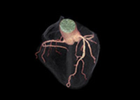
Studying tissue function with nuclear medicine aids in the diagnosis of many kinds of diseases, tumors, infection and other disorders in the brain, heart, lungs, bone, bowel, kidneys, thyroids and other internal structures. Nuclear cardiology focuses on imaging the heart to locate or diagnose problems with the heart. Scans may be performed in conjunction with other types of imaging (CT, MRI, X-ray) or with stress tests to determine the cause of unexplained chest pain.
PET (positron emission tomography) imaging is the most exciting new tool in diagnosing and staging cancer. PET studies the body's biological functions by measuring energy emitted by a radioactive substance injected prior to the procedure. The position and concentration of the injected material provides information about cell biochemistry and metabolism that can help diagnose a variety of diseases and other problems before physical damage has occurred. PET/CT combines the best of PET imaging with the best of CT imaging, allowing for a study of cell biochemistry and metabolism as well as an accurate localization of the area of concern.
First a radioactive material called a tracer is injected into the body. After enough time has passed for the tracer to spread throughout the body and settle in appropriate areas, the patient lies down on the PET/CT scan table and is moved into the machine, where an array of ring-shaped detectors create an image. These images appear on a computer screen beside the machine. The preliminary administering of the tracer takes half an hour to an hour to be absorbed. Scanning itself lasts 30-45 minutes.
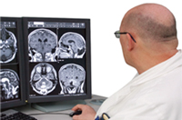
 Computed tomography, or CT (CAT) scan, is a sophisticated X-ray imaging system that scans thin "slices" of the body on all sides, then combines those slices into a highly detailed, three-dimensional digital image. The procedure is non-invasive, requires minimal radiation exposure and can simultaneously depict tissues of different densities, which is not possible with traditional x-ray methods. CT scans are highly useful for detecting problems, examining an injury, guiding biopsy needles and aiding in surgical preparation of almost all parts of the body.
Whereas traditional CT scans obtain one image per second, the ultrafast spiral CT technology available at Regional Radiology can produce eight images in the same amount of time. This means that scans of significant body areas and organs can be completed while the patient holds his or her breath. The rapidity of ultrafast CT is of major benefit to pediatric patients as well as those who are ill, claustrophobic or find it difficult to lie still for other reasons.
During the procedure, the patient is positioned on the table on his or her back, side or stomach, and may be provided with pillows for comfortable support. The technician leaves the room and the table moves slowly, often undetectably, through the CT scanner "doughnut." The X-ray beam inside the CT unit spirals slowly around the patient on all sides, creating 360-degree images or slices of the area being examined; as the table and the patient move through the unit, many slices are captured.
Sometimes an I.V. contrast is required for better visualization. Regional Radiology uses the safest non-ionic I.V. contrast on the market. Our team of I.V.-certified registered nurses monitors all contrast injections.
Ultrasound or sonography produces real-time, multi-dimensional images of the body's internal organs and tissues by mapping the reflections or "echoes" of high-frequency sound waves. Ultrasound is especially useful for examining the bladder, uterus, ovaries, prostate, testicles and scrotum, and for obtaining images of fetuses in the womb. In addition, ultrasound is often used as a real-time guide during needle biopsies for the precise sampling of tissue. During the procedure, the patient is positioned on an examination table and a warm, clear gel is applied to the area being examined. The sonographer or radiologist then presses a transducer firmly against the skin and sweeps it back and forth to obtain the image. In a transvaginal or transrectal ultrasound, a thin wand-like transducer will be covered, lubricated and placed in the vagina or rectum. 3-D ultrasound produces images in three dimensions, which is often of particular interest to obstetric patients for fetal imaging.
3-D ultrasound sonohysterography is used to detect polyps, fibroids, cancer or other anomalies of the female reproductive system by injecting a sterile saline (salt-water) solution into the uterus and performing an ultrasound examination. Patients suffering from infertility may be tested for patency of the fallopian tubes by injecting air along with the saline into the uterus.
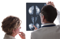 Mammography is an X-ray exam used specifically to obtain images of the breast. It is a highly useful tool in the early detection of breast cancer because it may show an abnormality in breast tissue before the patient or doctor can feel it. Screening and diagnostic mammography can aid in the detection and diagnosis of breast diseases, lumps, cysts and benign and malignant tumors. During the procedure, the breast is placed on a small platform and compressed with a paddle while it is exposed to a small amount of radiation. At Regional Radiology, images are displayed on a computer screen. Mammograms are recommended for patients who are at increased risk of developing breast cancer and other diseases, including yearly exams for women over 40.
Abnormalities discovered in the breast may be tested and treated with surgical or non-surgical (needle) biopsy. Needle biopsies are performed using several methods and with different specialized equipment depending on the type of abnormality.
 Physicians may use DEXA to measure bone density to detect osteoporosis and other diseases, to follow the progress of treatments for such conditions, and to gauge the patient's risk of developing bone fractures. DEXA (Dual-Energy X-ray Absorptiometry) is an enhanced X-ray image of the skeletal system that provides the most accurate measurements of bone density available. During the procedure, a beam of low-dose X-rays is targeted at the area, usually the lower spine and hips. The energy is measured as it is absorbed by soft tissue and by bone, and special computer software then processes the gathered information. Exams are painless and take 10-30 minutes.
In an upper GI series or barium swallow exam, X-ray images are taken of the esophagus, stomach and small intestine after the patient ingests a milkshake-like drink containing barium. The test takes three to six hours as the barium contrast travels through the digestive system and multiple X-rays are taken of the patient in various positions.
A barium enema is an X-ray examination of the large intestine. During the procedure, the patient lies on a table while liquid barium is delivered into the colon through a well-lubricated enema tube in the rectum. X-ray images are taken of the patient in various positions. A balloon may be inserted to keep the barium in place, and air may be used to inflate the colon and enhance the image. The tube is then removed and the patient expels as much of the barium as he or she can. A few more X-rays may be taken when the barium has been expelled.
An I.V.P. or intravenous pyelogram is an X-ray examination of the kidneys, bladder and uterus. Images are taken after a contrast dye containing iodine is injected through a vein and travels to the kidneys. The exam takes about one hour. The patient is usually asked to urinate before and immediately after the procedure.
Our system features three magnets:
CT imaging is performed with an ultrafast GE CT at our Bard Avenue and Richmond Avenue sites and with a Siemens Multi-detector CT at our Outerbridge location.
Ultrafast CT imaging is performed on GE Spiral CT equipment.
 © 2021 Regional Radiology and Advice Media. All Rights Reserved.
Powered by Advice Media.
