
Regional Radiation Oncology is one of only a few radiation oncology practices in the country to offer a designated PET/CT simulation in the department. Unlike traditional CT or fluoroscopic simulations, PET/CT simulation enables radiation oncologists to use information on both the tumor's location and activity to precisely pinpoint the radiation to only where it's needed. PET/CT simulation differs from a conventional PET/CT scan by the use of a special immobilization device that's necessary for radiation treatment.
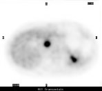
The PET/CT scan can provide the information of a PET and a CT scan. Merging the data together provides the additional advantage of fusion so that irregularities on either scan can be characterized. Most PET scans today are performed with an imaging radiopharmaceutical that acts like a very small amount of a sugar when it is administered intravenously. The imaging radiopharmaceutical most commonly used is FDG. FDG detects tissues that are under-using or over-using glucose. The abnormal uptake can be shown on the pictures. Cancer cells, because they are dividing faster than normal cells, tend to use more glucose than normal tissues. The PET scanner shows the distribution of the FDG in the body. The Food and Drug Administration (FDA) has found that FDG is indicated:
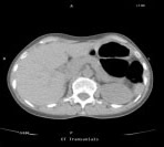
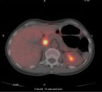
The CT scan provides detailed information about the structures in the body. The CT scan can show the dimension of vessels, lymph nodes and organ systems, showing changes in size, location, density and more. The merging of the two data sets- glucose metabolism and structure-provides the precise co-localization of anatomy and function.
 |
 |
 |
||||
| On a CT image, the physician sees the body's anatomic structure. | On a PET image, the physician sees how the body is functioning. | When combined into a PET/CT image, the patient's complete picture is revealed. |
Regional Radiology is now performing a new PET/CT bone scan using a fluoride agent F-18 NaF which provides more sensitivity and specificity for diagnosing bone metastasis. Instead of a conventional bone scan this test utilizing NaF offers higher contrast and using PET offers better spatial resolution……all providing a better image in detecting smaller bone lesions. Reimbursement for this new test is covered only through Medicare at this time. Please call Regional Radiology regarding NaF and PET/CT imaging questions.
 Your PET exam results may have a major impact on your physician's diagnosis of a potential health problem - and, should a disease be detected, how your return to health is managed.
A PET study not only helps your physician diagnose a problem; it also helps your physician predict the likely outcome of various therapeutic alternatives, pinpoint the best approach to treatment, and monitor your progress. If you're not responding as well as expected, you can be switched to a more effective therapy immediately.
Just ask your physician what he or she hopes to learn from your PET exam.
After reviewing your history and any prior exam, you'll receive a radiopharmaceutical injection. This is a radioactive tracer that must pass multiple quality control measures before it is used for any patient injection.
For most studies, you'll have to wait for the radiopharmaceutical to distribute itself - typically from 30 minutes to an hour. You may be able to read, speak or listen to music until your scan begins - and perhaps during the scan itself. However, if your brain is being scanned, you will be asked to wait in a quiet, dimly lit room, without stimulating your brain by reading or talking.
If you're having a heart study, on the other hand, you may not have to wait at all; the radiopharmaceuticals used for cardiac exams are often administered just before scanning begins.
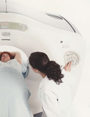
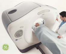
When you're ready for scanning, you'll lie on a comfortable table that moves slowly through the ring-like PET scanner as it acquires the information it needs to generate diagnostic images. You will be asked to lie very still, because movement can interfere with the results.
You shouldn't feel a thing during the scan, which can last anywhere from 15 to 60 minutes. Then, unless the physician sees a need for acquiring additional information, you will be free to leave.
 Your exam will vary depending on what your physician is looking for, and what is discovered along the way. Expect to spend two to three hours getting your PET exam.
You may leave as soon as the scan is complete. Unless you've received special instructions, you'll be able to eat and drink immediately - drinking lots of fluids will help remove any of the radiopharmaceutical that may still be in your system.
In the meantime, your results will be prepared for review and the findings forwarded to your physician, who will tell you what has been learned.
A PET, PET/CT study is similar to many other diagnostic procedures, from CT and MRI to Nuclear Medicine. Although the radiation you receive is different, it's roughly equivalent to what you'd receive from other diagnostic imaging exams such as CT (a couple of chest X-rays).
Radiopharmaceuticals used in PET don't remain in your system long, so there's no reason to avoid interacting with other people once you've left. Patients may want to avoid direct contact with an infant or pregnant woman for a few hours after the injection.
Please consult your physician with any additional questions or concerns.
 © 2021 Regional Radiology and Advice Media. All Rights Reserved.
Powered by Advice Media.
