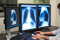
 Regional Radiology now provides digital Radiography exclusively. This allows us to process images faster and provides the radiologists with the ability to manipulate images to enhance contrast and definition. These features allow the radiologist to better evaluate areas of concern and many times alleviates the need for repeat films and additional exposure to the patient. Additionally, digital films are available for immediate viewing by the referring physician through use of our web server.
Fluoroscopy is used to obtain "live" X-ray images of a patient. During the procedure, x-rays pass through the body area being imaged, where they strike a fluorescent plate and are transmitted to a computer. The radiologist can then see the images on the computer screen in real-time.
Hysterosalpingography or HSG is an X-ray exam of the uterus and fallopian tubes using a contrast injected through the vagina. The test is usually performed to detect abnormalities or blockages of the reproductive organs in women suffering from infertility.
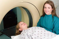 MRI stands for Magnetic Resonance Imaging, a non-invasive, radiation-free scanning technology that uses radio waves and magnetic fields to produce clear and detailed three-dimensional images of the body. MRI can be used to identify or precisely locate an injury or abnormality, to scan for developing problems or analyze damage from previous trauma, and to aid in the planning of surgery. MRI produces images of any area of the body and can be an invaluable tool for detecting tumors, infection, cancer and damage to the eye and inner ear, nervous system, heart and blood vessels, joint and musculoskeletal systems, major organs and male and female reproductive systems.
Unlike x-rays, radioisotopes and CT scanning, MRI uses radiofrequency waves, making it safer than other methods that use radiation. Radio waves detect differences in water concentration and distribution in various body tissues. Total exam time can be between 30 to 45 minutes. Because MRI requires the use of magnetic fields, patients will be asked to remove all metal objects. You must also be sure to make the MR staff aware of any implants or devices with a metal component prior to the exam.
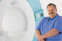 You need an MRI but you are anxious or claustrophobic or may be a little large than normal and haven't been able to fit in the bore. This used to be a problem. Not anymore! At Regional Radiology we offer a high field open MRI with an open bore design. Not all open MRI's offer the same technology. With the older open designs there was a trade off in image quality and speed. Most open machines do not offer a high magnetic field. The strength of the magnetic field determines the quality of the pictures. At Regional Radiology our new open bore Espree offers a 1.5 tesla magnetic field with the advantage of an open design. This makes it appropriate for people who suffer with claustrophobia, for children and the elderly who may need some hand holding and for the obese patient. The new design is faster and quieter than the older design making it more patient friendly with no trade off in image quality.
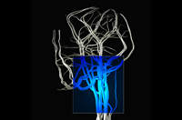 MR angiography, or MRA, is an MRI study of blood vessels using radio waves without the need for contrast material. It is used to detect, diagnose and aid in the treatment of stroke, aneurysms, and vascular disease in the heart, head, major organs and extremities. The procedure is painless, entails short examination and recovery times, and takes the place of catheter angiography and exploratory surgery so there is no risk of damaging an artery.
MR Enterography (MR-E) is a new imaging exam for evaluating Crohn's disease, now available at Regional Radiology. Crohn’s disease is a chronic condition which can strike early in life, requiring patients to undergo multiple scans over their lifetimes. There is a growing awareness of the risks of radiation exposure in these young patients. Our high field 1.5-Tesla MRI allows us to image the small and large bowel in great detail, without radiation exposure. If you are a patient with Crohn’s disease, MR Enterography may reduce your lifetime exposure to radiation.
MR-Enterography is available to all age groups. Ask your physician if you are a candidate for this procedure. A prescription from your physician is required. Call 718-605-6500 to schedule your MR-E appointment.
Nuclear medicine images body functions based on energy emitted by a radioactive substance ingested prior to examination. Involving only a small dose of radiation (about equivalent to the amount in a regular X-ray) that carries near zero risk of allergic reaction, nuclear medicine is a non-invasive alternative to exploratory surgery. The main drawback is the amount of time needed for each test -- absorption of the radioactive material can take anywhere from a few hours to a few days, and the scan itself can last as long as three hours.
First, the radioactive material (called a tracer) required for imaging is administered orally or by intravenous injection. Once inside the body, the tracer spreads, collects in certain areas and reacts with body tissue to produce gamma radiation. After sufficient time has elapsed for full absorption of the tracer, the patient lies down on the scanning table and is placed beneath or within the scanning device. Cameras detect the emitted gamma radiation, and a computer processes and displays the images.
Neither the injected tracer nor the scans themselves cause any pain, although there may be some discomfort associated with the actual injection or having to lie still during the scan, or (if one is necessary) with a catheter placed in the bladder.
 © 2021 Regional Radiology and Advice Media. All Rights Reserved.
Powered by Advice Media.
